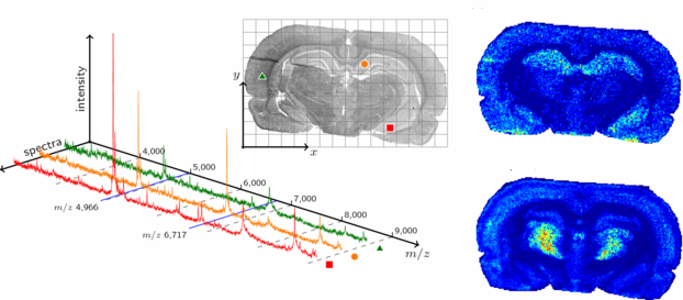
Mass spectrometry imaging (MSI) is a new method in pathology to study proteomics in tissues at an unprecedented spatial resolution. Whereas conventional histopathology typically uses immunohistochemistry staining against a few molecular to visualize the presence of a few biomarkers, MSI allows a detailed spatial inspection of the molecular composition of a tissue in terms of peptides and protein, and correlate this with histological images. This technique holds important promises for biomedical research into e.g. biomarker discovery as well as for clinical diagnosis.
Together with the molecular tumor diagnostics unit from the National Center for Tumor Diseases (NCT Dresden) at the Institute of Pathology, we are developing new bioinformatics and bioimaging methods, based on a variety of machine learning methods, to analyze mass spec images for diagnostic purposes.
Specifically, based on a growing clinical dataset from MALDI (matrix-assisted laser desorption ionization) imaging of common and rare tumor types, we develop methods to
- identify and characterize various tumor types according to their mass-charge (m/z) profiles using cluster analysis, and
- localize and accurately segment tumor tissue regions using convolutional neural networks.
collaborators
- Dr. Falk Zakrzewski, Institute of Pathology, University Hospital Dresden
- Dr. med. Pia Hönscheid, Molecular Tumor Diagnostics Unit, National Center for Tumor Diseases (NCT Dresden).